

Pleural fluid smear
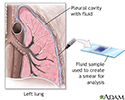
Pleural fluid smear is a lab test to check for bacteria, fungi, mycobacteria, or abnormal cells in a sample of the fluid that has collected in the pleural space. This is the space between the lining of the outside of the lungs (pleura) and the wall of the chest. When fluid collects in the pleural space, the condition is called pleural effusion.

A procedure called thoracentesis is used to get a sample of pleural fluid. Your health care provider sends the fluid to a lab where a technician examines a sample of pleural fluid under the microscope. If bacteria, mycobacteria, or fungi are found, other methods may be used to further identify those organisms.
How to Prepare for the Test
No special preparation is needed before the test. An ultrasound, chest CT scan, or chest x-ray will be performed before and after the test.


Do not cough, breathe deeply, or move during the test to avoid injury to the lung.
Tell your provider if you take medicines to thin the blood.
How the Test will Feel
For thoracentesis, you sit on the edge of a chair or bed with your head and arms resting on a table. Your provider cleans your skin around the insertion site. Numbing medicine (anesthetic) is injected into your skin.
A needle is placed through the skin and muscles of your chest wall into the space around the lungs, called the pleural space. As fluid drains into a collection bottle, you may cough a bit. This is because your lung re-expands to fill the space where fluid had been. This sensation may last for a few hours after the test.
During the test, tell your provider if you have sharp chest pain or shortness of breath.
Ultrasound is often used to decide where to insert the needle and to get a better view of the fluid in your chest.
Why the Test is Performed
The test is performed if you have a pleural effusion and its cause is not known, especially if your provider suspects an infection or cancer.

Normal Results
Normally, no bacteria, fungi, or cancer cells are present in the pleural fluid.
Normal value ranges may vary slightly among different labs. Talk to your provider about the meaning of your specific test results.
What Abnormal Results Mean
Positive results may indicate that infection, or cancer cells, are present. Other tests can help identify the specific type of infection or cancer. Sometimes, the test may show abnormalities (such as special types of cells) from conditions such as systemic lupus erythematosus.

Risks
Risks of thoracentesis are:
- Collapsed lung (pneumothorax)
- Excessive loss of blood
- Fluid re-accumulation
- Infection
- Pulmonary edema (swelling in the lungs)
- Respiratory distress
- Cough that doesn't go away
References
Broaddus VC, Light RW. Pleural effusion. In: Broaddus VC, Ernst JD, King TE, et al, eds. Murray and Nadel's Textbook of Respiratory Medicine. 7th ed. Philadelphia, PA: Elsevier; 2022:chap 108.
McCool FD. Diseases of the diaphragm, chest wall, pleura, and mediastinum. In: Goldman L, Cooney KA, eds. Goldman-Cecil Medicine. 27th ed. Philadelphia, PA: Elsevier; 2024:chap 86.
Ruhl TS, Good JL. Thoracentesis. In: Fowler GC, ed. Pfenninger and Fowler's Procedures for Primary Care. 4th ed. Philadelphia, PA: Elsevier; 2020:chap 218.
Review Date: 11/6/2025
Reviewed By: Jacob Berman, MD, MPH, Associate Professor of Medicine, Division of General Internal Medicine, University of Washington School of Medicine, Seattle, WA. Also reviewed by David C. Dugdale, MD, Medical Director, Brenda Conaway, Editorial Director, and the A.D.A.M. Editorial team.
© 1997- A.D.A.M., a business unit of Ebix, Inc. Any duplication or distribution of the information contained herein is strictly prohibited.
 All rights reserved.