

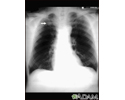
Chest x-ray
Chest radiography; Serial chest x-ray; X-ray - chest
A chest x-ray is an x-ray of the chest, lungs, heart, large arteries, ribs, and diaphragm.

How the Test is Performed
You stand in front of the x-ray machine. You will be told to take a breath in and hold it when the x-ray is taken.
Two images are usually taken. You will first need to stand facing the machine, and then sideways.
How to Prepare for the Test
Tell your health care provider if you are pregnant. Chest x-rays are generally avoided during pregnancy, and special precautions are taken if they are needed.
How the Test will Feel
There is no discomfort. The imaging plate may feel cold.
Why the Test is Performed
Your provider may order a chest x-ray if you have any of the following symptoms:
- A persistent cough
- Chest pain from a chest injury (with a possible rib fracture or lung complication) or from heart problems
- Coughing up blood
- Difficulty breathing
- Fever


It may also be done if you have signs of tuberculosis, lung cancer, or other chest or lung diseases.


A serial chest x-ray is one that is repeated. It may be done to monitor changes found on a past chest x-ray.
What Abnormal Results Mean
Abnormal results may be due to many things, including:
In the lungs:
- Collapsed lung
- Collection of fluid around the lung
- Lung tumor (noncancerous or cancerous)
- Malformation of the blood vessels
- Pneumonia
- Scarring of lung tissue
- Tuberculosis
- Atelectasis

In the heart:
- Problems with the size, position or shape of the heart
- Problems with the position, size and shape of the large arteries
- Evidence of heart failure
In the bones:
- Fractures or other problems of the ribs and spine
- Osteoporosis

In the mediastinum (middle part of the chest):
- Enlargement, which might be related to infection or tumor
Risks
There is low radiation exposure. X-rays are monitored and regulated to provide the minimum amount of radiation exposure needed to produce the image. Most experts feel that the benefits outweigh the risks. Pregnant women and children are more sensitive to the risks of x-rays.
References
Felker GM, Teerlink JR. Diagnosis and management of acute heart failure. In: Libby P, Bonow RO, Mann DL, Tomaselli GF, Bhatt DL, Solomon SD, eds. Braunwald's Heart Disease: A Textbook of Cardiovascular Medicine. 12th ed. Philadelphia, PA: Elsevier; 2022:chap 49.
Jokerst CE, Gotway MB. Thoracic radiology: noninvasive diagnostic imaging. In: Broaddus VC, Ernst JD, King TE, et al, eds. Murray and Nadel's Textbook of Respiratory Medicine. 7th ed. Philadelphia, PA: Elsevier; 2022:chap 20.
Nair A, Barnett JL, Semple TR. Current status of thoracic imaging. In: Adam A, Dixon AK, Gillard JH, Schaefer-Prokop CM, eds. Grainger & Allison's Diagnostic Radiology. 7th ed. Philadelphia, PA: Elsevier; 2021:chap 1.
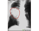
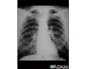
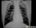
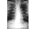
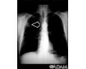
Aortic rupture - chest X-ray - illustration
Aortic rupture - chest X-ray
illustration
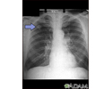
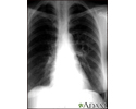
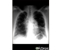
Lung cancer - frontal chest X-ray - illustration
Lung cancer - frontal chest X-ray
illustration
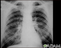
Adenocarcinoma - chest x-ray - illustration
Adenocarcinoma - chest x-ray
illustration
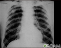
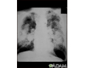
Coal worker's lungs - chest x-ray - illustration
Coal worker's lungs - chest x-ray
illustration
Coccidioidomycosis - chest X-ray - illustration
Coccidioidomycosis - chest X-ray
illustration
Coal workers pneumoconiosis - stage II - illustration
Coal workers pneumoconiosis - stage II
illustration
Coal workers pneumoconiosis - stage II - illustration
Coal workers pneumoconiosis - stage II
illustration
Coal workers pneumoconiosis, complicated - illustration
Coal workers pneumoconiosis, complicated
illustration
Coal workers pneumoconiosis, complicated - illustration
Coal workers pneumoconiosis, complicated
illustration
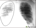
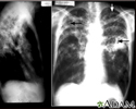
Tuberculosis, advanced - chest X-rays - illustration
Tuberculosis, advanced - chest X-rays
illustration
Pulmonary nodule - front view chest x-ray - illustration
Pulmonary nodule - front view chest x-ray
illustration
Sarcoid, stage II - chest X-ray - illustration
Sarcoid, stage II - chest X-ray
illustration
Sarcoid, stage IV - chest x-ray - illustration
Sarcoid, stage IV - chest x-ray
illustration
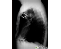
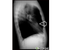
Pulmonary mass - side view chest X-ray - illustration
Pulmonary mass - side view chest X-ray
illustration
Bronchial cancer - chest X-ray - illustration
Bronchial cancer - chest X-ray
illustration
Lung nodule, right middle lobe - chest X-ray - illustration
Lung nodule, right middle lobe - chest X-ray
illustration
Lung mass, right upper lung - chest X-ray - illustration
Lung mass, right upper lung - chest X-ray
illustration
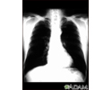
Lung nodule - front view chest X-ray - illustration
Lung nodule - front view chest X-ray
illustration
Aortic rupture - chest X-ray - illustration
Aortic rupture - chest X-ray
illustration
Lung cancer - frontal chest X-ray - illustration
Lung cancer - frontal chest X-ray
illustration
Adenocarcinoma - chest x-ray - illustration
Adenocarcinoma - chest x-ray
illustration
Coal worker's lungs - chest x-ray - illustration
Coal worker's lungs - chest x-ray
illustration
Coccidioidomycosis - chest X-ray - illustration
Coccidioidomycosis - chest X-ray
illustration
Coal workers pneumoconiosis - stage II - illustration
Coal workers pneumoconiosis - stage II
illustration
Coal workers pneumoconiosis - stage II - illustration
Coal workers pneumoconiosis - stage II
illustration
Coal workers pneumoconiosis, complicated - illustration
Coal workers pneumoconiosis, complicated
illustration
Coal workers pneumoconiosis, complicated - illustration
Coal workers pneumoconiosis, complicated
illustration
Tuberculosis, advanced - chest X-rays - illustration
Tuberculosis, advanced - chest X-rays
illustration
Pulmonary nodule - front view chest x-ray - illustration
Pulmonary nodule - front view chest x-ray
illustration
Sarcoid, stage II - chest X-ray - illustration
Sarcoid, stage II - chest X-ray
illustration
Sarcoid, stage IV - chest x-ray - illustration
Sarcoid, stage IV - chest x-ray
illustration
Pulmonary mass - side view chest X-ray - illustration
Pulmonary mass - side view chest X-ray
illustration
Bronchial cancer - chest X-ray - illustration
Bronchial cancer - chest X-ray
illustration
Lung nodule, right middle lobe - chest X-ray - illustration
Lung nodule, right middle lobe - chest X-ray
illustration
Lung mass, right upper lung - chest X-ray - illustration
Lung mass, right upper lung - chest X-ray
illustration
Lung nodule - front view chest X-ray - illustration
Lung nodule - front view chest X-ray
illustration
Review Date: 8/19/2024
Reviewed By: Allen J. Blaivas, DO, Division of Pulmonary, Critical Care, and Sleep Medicine, VA New Jersey Health Care System, Clinical Assistant Professor, Rutgers New Jersey Medical School, East Orange, NJ. Review provided by VeriMed Healthcare Network. Also reviewed by David C. Dugdale, MD, Medical Director, Brenda Conaway, Editorial Director, and the A.D.A.M. Editorial team.
 All rights reserved.