

Ultrasound
Sonogram
Ultrasound uses high-frequency sound waves to make images of organs and structures inside the body.
How the Test is Performed
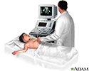
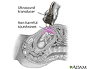
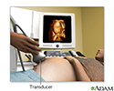
An ultrasound machine makes images so that organs inside the body can be examined. The machine sends out high-frequency sound waves, which reflect off body structures. A computer receives the waves and uses them to create a picture. Unlike with an x-ray or CT scan, this test does not use ionizing radiation.


The test is done in the ultrasound or radiology department.
- You will lie down for the test.
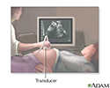
- A clear, water-based gel is applied to the skin on the area to be examined. The gel helps with the transmission of the sound waves.
- A handheld probe called a transducer is moved over the area being examined. You may need to change position so that other areas can be examined.
How to Prepare for the Test
Your preparation will depend on the part of the body being examined.
How the Test will Feel
Most of the time, ultrasound procedures do not cause discomfort. The conducting gel may feel a little cold and wet. You will feel the sonographer press the ultrasound probe against your body in the area they are reviewing.
Why the Test is Performed
The reason for the test will depend on your symptoms. An ultrasound test may be used to identify problems involving:
- Arteries in the neck
- Veins or arteries in the arms, legs, or abdomen
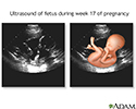
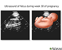
- Pregnancy
- Pelvis
- Abdomen and kidneys
- Breast
- Thyroid
- Eye and orbit


Normal Results
Results are considered normal if the organs and structures being examined look OK.
What Abnormal Results Mean
The meaning of abnormal results will depend on the part of the body being examined and the problem found. Talk to your health care provider about your questions and concerns.
Risks
There are no known risks. The test does not use ionizing radiation.
Considerations
Some types of ultrasound tests need to be done with a probe that is inserted into your body. Talk to your provider about how your test will be done.
References
Butts C. Ultrasound. In: Roberts JR, Custalow CB, Thomsen TW, eds. Roberts and Hedges' Clinical Procedures in Emergency Medicine and Acute Care. 7th ed. Philadelphia, PA: Elsevier; 2019:chap 66.
Fowler GC, Lefevre N. Emergency department, hospitalist, and office ultrasound (POCUS). In: Fowler GC, ed. Pfenninger and Fowler's Procedures for Primary Care. 4th ed. Philadelphia, PA: Elsevier; 2020:chap 214.
Morris AE, Adamson R, Frank J. Ultrasonography: principles and basic thoracic and vascular imaging. In: Broaddus VC, Ernst JD, King TE, et al, eds. Murray and Nadel's Textbook of Respiratory Medicine. 7th ed. Philadelphia, PA: Elsevier; 2022:chap 23.
Zhang D, Kahlili K, Yu H, Levine D. Physics of ultrasound. In: Rumack CM, Levine D, eds. Diagnostic Ultrasound. 6th ed. Philadelphia, PA: Elsevier; 2024:chap 1.
Abdominal ultrasound - illustration
Abdominal ultrasound
illustration
Ultrasound in pregnancy - illustration
Ultrasound in pregnancy
illustration
17 week ultrasound - illustration
17 week ultrasound
illustration
30 week ultrasound - illustration
30 week ultrasound
illustration
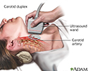
Carotid duplex - illustration
Carotid duplex
illustration
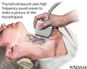
Thyroid ultrasound - illustration
Thyroid ultrasound
illustration
Ultrasound - illustration
Ultrasound
illustration
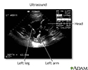
Ultrasound, normal fetus - ventricles of brain - illustration
Ultrasound, normal fetus - ventricles of brain
illustration
3D ultrasound - illustration
3D ultrasound
illustration
Ultrasound comparison - illustration
Ultrasound comparison
illustration
Abdominal ultrasound - illustration
Abdominal ultrasound
illustration
Ultrasound in pregnancy - illustration
Ultrasound in pregnancy
illustration
17 week ultrasound - illustration
17 week ultrasound
illustration
30 week ultrasound - illustration
30 week ultrasound
illustration
Carotid duplex - illustration
Carotid duplex
illustration
Thyroid ultrasound - illustration
Thyroid ultrasound
illustration
Ultrasound - illustration
Ultrasound
illustration
Ultrasound, normal fetus - ventricles of brain - illustration
Ultrasound, normal fetus - ventricles of brain
illustration
3D ultrasound - illustration
3D ultrasound
illustration
Ultrasound comparison - illustration
Ultrasound comparison
illustration
Review Date: 7/15/2024
Reviewed By: Jason Levy, MD, FSIR, Northside Radiology Associates, Atlanta, GA. Also reviewed by David C. Dugdale, MD, Medical Director, Brenda Conaway, Editorial Director, and the A.D.A.M. Editorial team.
 All rights reserved.