

Tetralogy of Fallot
Tet; TOF; Congenital heart defect - tetralogy; Cyanotic heart disease - tetralogy; Birth defect - tetralogy
Tetralogy of Fallot is a type of congenital heart defect. Congenital means that it is present at birth.
Causes
Tetralogy of Fallot causes low oxygen levels in the blood. This leads to cyanosis (a bluish-purple color to the skin).
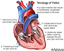
The classic form includes four defects of the heart and its major blood vessels:
- Ventricular septal defect (hole between the right and left ventricles)
- Narrowing of the pulmonary outflow tract (the valve and artery that connect the heart with the lungs)
- Overriding aorta (the artery that carries oxygen-rich blood to the body) that is shifted over the right ventricle and ventricular septal defect, instead of coming out only from the left ventricle
- Thickened wall of the right ventricle (right ventricular hypertrophy)

Tetralogy of Fallot is rare, but it is the most common form of cyanotic congenital heart disease. It occurs equally as often in males and females. People with tetralogy of Fallot are more likely to also have other congenital defects.
The cause of most congenital heart defects is unknown. Many factors seem to be involved.
Factors that increase the risk for this condition during pregnancy include:
- Alcoholism in the mother
- Diabetes
- Mother who is over 40 years old
- Poor nutrition during pregnancy
- Rubella or other viral illnesses during pregnancy

Children with tetralogy of Fallot are more likely to have chromosome disorders, such as Down syndrome, Alagille syndrome, and DiGeorge syndrome (a condition that causes heart defects, low calcium levels, and poor immune function).
Symptoms
Symptoms include:
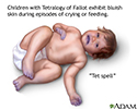
- Bluish color to the skin (cyanosis) due to low oxygen level in the blood, which gets worse when the baby is upset
- Clubbing of fingers (skin or bone enlargement around the fingernails)
- Difficulty feeding (poor feeding habits)
- Failure to gain weight
- Passing out
- Poor development
- Squatting during episodes of cyanosis (which increases blood flow to the lungs)


Exams and Tests
A physical exam with a stethoscope almost always reveals a heart murmur.
Tests may include:
- Chest x-ray
- Complete blood count (CBC)
- Echocardiogram
- Electrocardiogram (ECG)
- MRI of the heart (generally after surgery)
- CT of the heart


Treatment
Surgery to repair tetralogy of Fallot is done when the infant is very young, typically before 6 months of age. Sometimes, more than one surgery is needed. When more than one surgery is used, the first surgery is done to help increase blood flow to the lungs.
Surgery to correct the problem may be done at a later time. Often only one corrective surgery is performed in the first few months of life. Corrective surgery is done to widen part of the narrowed pulmonary tract and close the ventricular septal defect with a patch.
Outlook (Prognosis)
Most cases can be corrected with surgery. Babies who have surgery usually do well. More than 90% survive to adulthood and live active, healthy, and productive lives. Without surgery, death often occurs by the time the person reaches age 20.
People who have excessive leakiness of the pulmonary valve after surgery (which is common) may need to have the valve replaced.
Regular follow-up with a cardiologist is strongly recommended.
Possible Complications
Complications may include:
- Delayed growth and development
- Irregular heart rhythms (arrhythmias)
- Seizures during periods when there is not enough oxygen
- Death from cardiac arrest, even after surgical repair


When to Contact a Medical Professional
Contact your child's health care provider if new unexplained symptoms develop or your child is having an episode of cyanosis (blue skin).
If a child with tetralogy of Fallot becomes blue, immediately place the child on their side or back and put the knees up to the chest. Calm the child and seek medical attention right away.
Prevention
There is no known way to prevent the condition.
Some inherited factors may play a role in congenital heart disease. Many family members may be affected. If you are planning to get pregnant, talk to your provider about screening for genetic diseases.
Women who plan to become pregnant should be immunized against rubella if they are not already immune. Rubella infection in a pregnant woman can cause congenital heart disease.
Women who are pregnant should get good prenatal care:
- Avoid alcohol and illegal drugs during pregnancy.
- Tell your provider that you are pregnant before taking any new medicines.
- Have a blood test early in your pregnancy to see if you are immune to rubella. If you are not immune, avoid any possible exposure to rubella and get vaccinated right after delivery.
- Pregnant women who have diabetes should try to get good control over their blood sugar level.

References
Bernstein D. Evaluation and screening of the infant or child with congenital heart disease. In: Kliegman RM, St. Geme JW, Blum NJ, et al, eds. Nelson Textbook of Pediatrics. 22nd ed. Philadelphia, PA: Elsevier; 2025:chap 474.
Kliegman RM, St. Geme JW, Blum NJ, et al. Cyanotic congenital heart disease: lesions associated with decreased pulmonary blood flow. In: Kliegman RM, St. Geme JW, Blum NJ, et al, eds. Nelson Textbook of Pediatrics. 22nd ed. Philadelphia, PA: Elsevier; 2025:chap 479.
Valente AM, Dorfman AL, Babu-Narayan SV, Kreiger EV. Congenital heart disease in the adolescent and adult. In: Libby P, Bonow RO, Mann DL, Tomaselli GF, Bhatt DL, Solomon SD, eds. Braunwald's Heart Disease: A Textbook of Cardiovascular Medicine. 12th ed. Philadelphia, PA: Elsevier; 2022:chap 82.
Well A, Fraser CD. Congenital heart disease. In: Townsend CM Jr, Beauchamp RD, Evers BM, Mattox KL, eds. Sabiston Textbook of Surgery. 21st ed. St Louis, MO: Elsevier; 2022:chap 59.
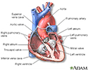
Heart - section through the middle - illustration
Heart - section through the middle
illustration
Tetralogy of Fallot - illustration
Tetralogy of Fallot
illustration
Cyanotic 'Tet spell' - illustration
Cyanotic 'Tet spell'
illustration
Review Date: 10/1/2025
Reviewed By: Thomas S. Metkus MD, PhD, Associate Professor of Medicine and Surgery, Johns Hopkins University School of Medicine, Baltimore, MD. Also reviewed by David C. Dugdale, MD, Medical Director, Brenda Conaway, Editorial Director, and the A.D.A.M. Editorial team.
© 1997- A.D.A.M., a business unit of Ebix, Inc. Any duplication or distribution of the information contained herein is strictly prohibited.
 All rights reserved.