Truncus arteriosus
Truncus
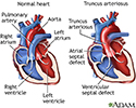
Truncus arteriosus is a rare type of heart disease in which a single blood vessel (truncus arteriosus) comes out of the right and left ventricles, instead of the normal 2 vessels (pulmonary artery and aorta). It is present at birth (congenital heart disease).
There are different types of truncus arteriosus.
Causes
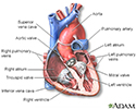
In normal circulation, the pulmonary artery comes out of the right ventricle and the aorta comes out of the left ventricle, which are separate from each other.
With truncus arteriosus, a single artery comes out of the ventricles. There is most often also a large hole between the 2 ventricles (ventricular septal defect). As a result, the blue (without oxygen) and red (oxygen-rich) blood mix.
Some of this mixed blood goes to the lungs, and some goes to the rest of the body. Often, more blood than usual ends up going to the lungs.
If this condition is not treated, two problems occur:
- Too much blood circulation in the lungs may cause extra fluid to build up in and around them. This makes it hard to breathe.
- If left untreated and more than normal blood flows to the lungs for a long time, the blood vessels to the lungs become permanently damaged. Over time, it becomes very hard for the heart to force blood through them. This is called pulmonary hypertension, which can be life threatening.
Symptoms
Symptoms include:
- Bluish skin (cyanosis)
- Delayed growth or growth failure
- Fatigue
- Lethargy
- Poor feeding
- Rapid breathing (tachypnea)
- Shortness of breath (dyspnea)
- Widening of the finger tips (clubbing)
Exams and Tests
A murmur is most often heard when listening to the heart with a stethoscope.
Tests include:
- ECG
- Echocardiogram
- Chest x-ray
- Cardiac catheterization
- MRI or CT scan of the heart
Treatment
Surgery is needed to treat this condition. The surgery creates 2 separate arteries.
In most cases, the truncal vessel is kept as the new aorta. A new pulmonary artery is created using tissue from another source or using a man-made tube. The branch pulmonary arteries are sewn to this new artery. The hole between the ventricles is closed.
Outlook (Prognosis)
Complete repair most often provides good results. Another procedure may be needed as the child grows, because the rebuilt pulmonary artery that uses tissue from another source will not grow with the child.
Untreated cases of truncus arteriosus result in death, often during the first year of life.
Possible Complications
Complications may include:
- Heart failure
- High blood pressure in the lungs (pulmonary hypertension)
When to Contact a Medical Professional
Call your health care provider if your infant or child:
- Appears lethargic
- Appears overly tired or mildly short of breath
- Does not eat well
- Does not seem to be growing or developing normally
If the skin, lips, or nail beds look blue or if the child seems to be very short of breath, take the child to the emergency room or have the child examined promptly.
Prevention
There is no known prevention. Early treatment can often prevent serious complications.
References
Valente AM, Dorfman AL, Babu-Narayan SV, Kreiger EV. Congenital heart disease in the adolescent and adult. In: Libby P, Bonow RO, Mann DL, Tomaselli GF, Bhatt DL, Solomon SD, eds. Braunwald's Heart Disease: A Textbook of Cardiovascular Medicine. 12th ed. Philadelphia, PA: Elsevier; 2022:chap 82.
Well A, Fraser CD. Congenital heart disease. In: Townsend CM Jr, Beauchamp RD, Evers BM, Mattox KL, eds. Sabiston Textbook of Surgery: The Biological Basis of Modern Surgical Practice. 21st ed. Philadelphia, PA: Elsevier; 2022:chap 59.
Heart - section through the middle - illustration
Heart - section through the middle
illustration
Truncus arteriosus - illustration
Truncus arteriosus
illustration
Review Date: 10/10/2021
Reviewed By: Michael A. Chen, MD, PhD, Associate Professor of Medicine, Division of Cardiology, Harborview Medical Center, University of Washington Medical School, Seattle, WA. Also reviewed by David Zieve, MD, MHA, Medical Director, Brenda Conaway, Editorial Director, and the A.D.A.M. Editorial team.


