

Aortopulmonary window
Aortopulmonary septal defect; Aortopulmonary fenestration; Congenital heart defect - aortopulmonary window; Birth defect heart - aortopulmonary window
Aortopulmonary window is a rare heart defect in which there is a hole connecting the major artery taking blood from the heart to the body (the aorta) and the one taking blood from the heart to the lungs (pulmonary artery). The condition is congenital, which means it is present at birth.
Causes
Normally, blood flows through the pulmonary artery into the lungs, where it picks up oxygen. Then the blood travels back to the heart and is pumped to the aorta and the rest of the body.
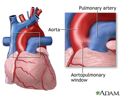
Babies with an aortopulmonary window have a hole in between the aorta and pulmonary artery. Because of this hole, blood from the aorta flows into the pulmonary artery, and as a result too much blood flows to the lungs. This causes high blood pressure in the lungs (a condition called pulmonary hypertension) and congestive heart failure. The bigger the defect, the more blood that is able to enter the pulmonary artery.
The condition occurs when the aorta and pulmonary artery do not divide normally as the baby develops in the womb.
Aortopulmonary window is very rare. It accounts for less than 1% of all congenital heart defects.
This condition can occur on its own or with other congenital heart defects such as:
- Tetralogy of Fallot
- Pulmonary atresia
- Truncus arteriosus
- Atrial septal defect
- Patent ductus arteriosus
- Interrupted aortic arch


Fifty percent of people usually have no other heart defects.
Symptoms
If the defect is small, it may not cause any symptoms. However, most defects are large.
Symptoms can include:
- Delayed growth
- Heart failure
- Irritability
- Poor eating and lack of weight gain
- Rapid breathing
- Rapid heartbeat
- Respiratory infections
Exams and Tests
The health care provider will usually hear an abnormal heart sound (murmur) when listening to the child's heart with a stethoscope.
The provider may order tests such as:
- Cardiac catheterization -- a thin tube inserted into blood vessels and/or arteries around the heart to view the heart and blood vessels and directly measure pressure in the heart and lungs.
- Chest x-ray.
- Echocardiogram.
- MRI of the heart.


Treatment
The condition usually requires open heart surgery to repair the defect. Surgery should be done as soon as possible after the diagnosis is made. In most cases, this is when the child is still a newborn.
During the procedure, a heart-lung machine takes over for the child's heart. The surgeon opens the aorta and closes the defect with a patch made either from a piece of the sac that encloses the heart (the pericardium) or a man-made material.
Outlook (Prognosis)
Surgery to correct an aortopulmonary window is successful in most cases. If the defect is treated quickly, the child should not have any lasting effects.
Possible Complications
Delaying treatment can lead to complications such as:
- Congestive heart failure
- Pulmonary hypertension or Eisenmenger syndrome
- Death

When to Contact a Medical Professional
Contact your child's provider if your child has symptoms of aortopulmonary window. The sooner this condition is diagnosed and treated, the better the child's prognosis.
Prevention
There is no known way to prevent aortopulmonary window.
References
Bernstein D. General principles of treatment of congenital heart disease. In: Kliegman RM, St. Geme JW, Blum NJ, et al, eds. Nelson Textbook of Pediatrics. 22nd ed. Philadelphia, PA: Elsevier; 2025:chap 483.
Gurvitz M, Krieger EV, Fuller S, et al. 2025 ACC/AHA/HRS/ISACHD/SCAI Guideline for the Management of Adults With Congenital Heart Disease: A Report of the American College of Cardiology/American Heart Association Joint Committee on Clinical Practice Guidelines. J Am Coll Cardiol. 2026;87(7):822-976. PMID: 41411480 pubmed.ncbi.nlm.nih.gov/41411480/.
Kliegman RM, St. Geme JW, Blum NJ, et al. Acyanotic congenital heart disease: left-to-right shunt lesions. In: Kliegman RM, St. Geme JW, Blum NJ, et al, eds. Nelson Textbook of Pediatrics. 22nd ed. Philadelphia, PA: Elsevier; 2025:chap 475.
Qureshi AM, Gowda ST, Justino H, Spicer DE, Anderson RH. Other malformations of the ventricular outflow tracts. In: Wernovsky G, Anderson RH, Kumar K, et al, eds. Anderson's Pediatric Cardiology. 4th ed. Philadelphia, PA: Elsevier; 2020:chap 51.
Krieger EV, Dorfman AL, Babu-Narayan SV, Valente AM. Congenital heart disease in the adolescent and adult. In: Bonow RO, Mann DL, Tomaselli GF, et al, eds. Braunwald's Heart Disease: A Textbook of Cardiovascular Medicine. 13th ed. Philadelphia, PA: Elsevier; 2026:chap 87.
Well A, Fraser CD. Congenital heart disease. In: Tyler DS, Hayes-Dixon A, Hines OJ, et al, eds. Sabiston Textbook of Surgery: The Biological Basis of Modern Surgical Practice. 22nd ed. Philadelphia, PA: Elsevier; 2026:chap 113.
Review Date: 2/9/2026
Reviewed By: Michael A. Chen, MD, PhD, Associate Professor of Medicine, Division of Cardiology, Harborview Medical Center, University of Washington Medical School, Seattle, WA. Also reviewed by David C. Dugdale, MD, Medical Director, Brenda Conaway, Editorial Director, and the A.D.A.M. Editorial team.
© 1997- A.D.A.M., a business unit of Ebix, Inc. Any duplication or distribution of the information contained herein is strictly prohibited.
 All rights reserved.