Calcium pyrophosphate arthritis
Calcium pyrophosphate dihydrate deposition disease; CPPD disease; Acute/chronic CPPD arthritis; Pseudogout; Pyrophosphate arthropathy; Chondrocalcinosis
Calcium pyrophosphate dihydrate (CPPD) arthritis is a joint disease that can cause attacks of arthritis. Like gout, crystals form in the joints. But in this arthritis, the crystals are not formed from uric acid.
Causes
Deposition of calcium pyrophosphate dihydrate (CPPD) causes this form of arthritis. The buildup of this chemical forms crystals in the cartilage of joints. This leads to attacks of joint swelling and pain in the knees, wrists, ankles, shoulders, and other joints. In contrast to gout, the metatarsal-phalangeal joint of the big toe is not affected.
Among older adults, CPPD is a common cause of sudden (acute) arthritis in one joint. The attack is caused by:
- Injury to the joint
- Medical stress
CPPD arthritis mainly affects the elderly because joint degeneration and osteoarthritis increases with age. Such joint damage increases the tendency of CPPD deposition. However, CPPD arthritis is more common in people with:
- Hemochromatosis
- Parathyroid disease
- Dialysis-dependent renal failure
Symptoms
In most cases, CPPD arthritis does not cause any symptoms. Instead, x-rays of affected joints such as knees show characteristic deposits of calcium.
Some people with chronic CPPD deposits in large joints may have the following symptoms:
- Pain
- Swelling
- Warmth
- Redness
Attacks of joint pain can last for months. There may be no symptoms between attacks.
In some people CPPD arthritis causes severe damage to a joint.
CPPD arthritis can also occur in the spine, both lower and upper. Pressure on spinal nerves may cause pain in the arms or legs.
Because the symptoms are similar, CPPD arthritis can be confused with:
- Gouty arthritis (gout)
- Osteoarthritis
- Rheumatoid arthritis
Exams and Tests
Most arthritic conditions show similar symptoms. Carefully testing the joint fluid for crystals can help the doctor detect the condition.
You may undergo the following tests:
- Joint fluid exam to detect white blood cells and calcium pyrophosphate crystals
- Joint x-rays to look for joint damage and calcium deposits in joint spaces
- Other joint imaging tests such as CT scan, MRI or ultrasound, if required
- Blood tests to screen for conditions that are linked to calcium pyrophosphate arthritis
Treatment
Treatment may involve removing fluid to relieve pressure in the joint. A needle is placed into the joint and fluid is aspirated. Some common treatment options are:
- Steroid injections: to treat severely swollen joints
- Oral steroids: to treat multiple swollen joints
- Nonsteroidal anti-inflammatory medicines (NSAIDs): to ease the pain
- Colchicine: to treat attacks of CPPD arthritis
- For chronic CPPD arthritis in multiple joints, methotrexate or hydroxychloroquine may be helpful
Outlook (Prognosis)
Most people do well with treatment to reduce the acute joint pain. A medicine such as colchicine may help prevent repeat attacks. There is no treatment to remove the CPPD crystals.
Possible Complications
Permanent joint damage can occur without treatment.
When to Contact a Medical Professional
Contact your health care provider if you have attacks of joint swelling and joint pain.
Prevention
There is no known way to prevent this disorder. However, treating other problems that may cause CPPD arthritis may make the condition less severe.
Regular follow-up visits may help prevent permanent damage of the affected joints.
References
Andrés M, Sivera F, Pascual E. Therapy for CPPD: options and evidence. Curr Rheumatol Rep. 2018;20(6):31. PMID: 29675606 pubmed.ncbi.nlm.nih.gov/29675606/.
Edwards NL. Crystal deposition diseases. In: Goldman L, Schafer AI, eds. Goldman-Cecil Medicine. 26th ed. Philadelphia, PA: Elsevier; 2020:chap 257.
Terkeltaub R. Calcium crystal disease: calcium pyrophosphate dihydrate and basic calcium phosphate. In: Firestein GS, Budd RC, Gabriel SE, Koretzky GA, McInnes IB, O'Dell JR, eds. Firestein & Kelley's Textbook of Rheumatology. 11th ed. Philadelphia, PA: Elsevier; 2021:chap 102.
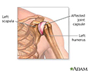
Shoulder joint inflammation - illustration
Shoulder joint inflammation
illustration
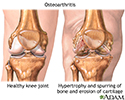
Osteoarthritis - illustration
Osteoarthritis
illustration
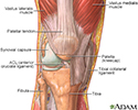
The structure of a joint - illustration
The structure of a joint
illustration
Review Date: 1/16/2022
Reviewed By: Diane M. Horowitz, MD, Rheumatology and Internal Medicine, Northwell Health, Great Neck, NY. Review provided by VeriMed Healthcare Network. Also reviewed by David Zieve, MD, MHA, Medical Director, Brenda Conaway, Editorial Director, and the A.D.A.M. Editorial team.


