

Health Library
Endometrial cancer
Endometrial adenocarcinoma; Uterine adenocarcinoma; Uterine cancer; Adenocarcinoma - endometrium; Adenocarcinoma - uterus; Cancer - uterine; Cancer - endometrial; Uterine corpus cancer
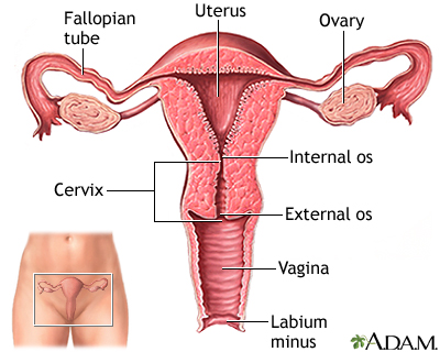
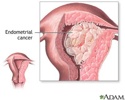
Endometrial cancer is cancer that starts in the endometrium, the lining of the uterus (womb).
Images
I Would Like to Learn About:
Causes
Endometrial cancer is the most common type of uterine cancer. The exact cause of endometrial cancer is not known. An increased level of estrogen hormone may play a role. This stimulates the buildup of the lining of the uterus. This can lead to abnormal overgrowth of the endometrium and cancer.
Most cases of endometrial cancer occur post menopause. The average age of diagnosis is around 60 years old.
The following factors related to your hormones increase your risk for endometrial cancer:
- Estrogen replacement therapy without the use of progesterone
- History of endometrial polyps
- Infrequent periods
- Never being pregnant
- Obesity
- Diabetes
- Polycystic ovary syndrome (PCOS)
- Starting menstruation at an early age (before age 12)
- Starting menopause after age 50
- Tamoxifen, a medicine used for breast cancer treatment
Women with the following conditions also seem to be at a higher risk for endometrial cancer:
- Colon or breast cancer
- Gallbladder disease
- High blood pressure
Symptoms
Symptoms of endometrial cancer include:
- Abnormal bleeding from the vagina, including bleeding between periods or spotting/bleeding after menopause
- Long, heavy, or frequent episodes of vaginal bleeding after age 40
- Lower abdominal pain or pelvic cramping
Exams and Tests
During the early stages of disease, a pelvic exam is often normal.
- In advanced stages, there may be changes in the size, shape, or feel of the uterus or surrounding structures.
- A Pap smear (may raise a suspicion for endometrial cancer, but does not diagnose it)
Based on your symptoms and other findings, other tests may be needed. Some can be done in your health care provider's office. Others may be done at a hospital or surgical center:
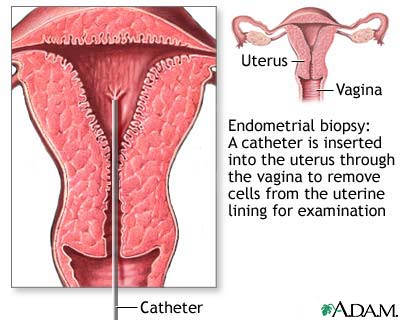
- Endometrial biopsy: Using a small thin catheter (tube), tissue is taken from the lining of the uterus (endometrium). The cells are examined under a microscope to see if any appear to be abnormal or cancerous.
- Hysteroscopy: A thin telescope-like device is inserted through the vagina and the opening of the cervix. It lets the provider view the inside of the uterus. Tissue can also be removed for analysis during the exam.
- Ultrasound: Sound waves are used to make a picture of the pelvic organs. The ultrasound may be performed abdominally or vaginally. An ultrasound can determine if the lining of the uterus appears abnormal or thickened.
- Sonohysterography: Fluid is placed in the uterus through a thin tube, while vaginal ultrasound images are made of the uterus. This procedure can be done to determine the presence of any abnormal uterine mass which may be an indication of cancer.
- Magnetic resonance imaging (MRI): In this imaging test, powerful magnets are used to create images of internal organs.
If cancer is found, imaging tests may be done to see if the cancer has spread to other parts of the body. This is called staging.
Stages of endometrial cancer are:
- Stage 1: The cancer is only in the uterus.
- Stage 2: The cancer is in the uterus and cervix.
- Stage 3: The cancer has spread outside of the uterus, but not beyond the true pelvis area. Cancer may involve the lymph nodes in the pelvis or near the aorta (the major artery in the abdomen).
- Stage 4: The cancer has spread to the inner surface of the bowel, bladder, abdomen, or other organs.
Cancer is also described as grade 1, 2, or 3. Grade 1 is the least aggressive, and grade 3 is the most aggressive. Aggressive means that the cancer grows and spreads quickly.
Treatment
Treatment options include:
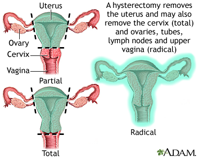
Surgery to remove the uterus (hysterectomy) may be done in women with stage 1 cancer. The surgeon may also remove the tubes and ovaries.
Surgery combined with radiation therapy is another treatment option. It is often used for women with:
- Stage 1 disease that has a high chance of returning or is a grade 2 or 3
- Stage 2 or 3 disease
Chemotherapy or hormonal therapy may be considered in some cases, most often for those with stage 3 and 4 disease.
Support Groups
You can ease the stress of illness by joining a cancer support group. Sharing with others who have common experiences and problems can help you not feel alone.
Outlook (Prognosis)
Endometrial cancer is usually diagnosed at an early stage.
If the cancer has not spread, 95% of women are alive 5 years after treatment. If the cancer has spread to distant organs, about 25% of women are still alive after 5 years.
Possible Complications
Complications may include any of the following:
- Anemia due to blood loss (before diagnosis)
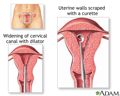
- Perforation (hole) of the uterus, which may occur during a dilation and curettage (D and C) or endometrial biopsy
- Problems from surgery, radiation, and chemotherapy
When to Contact a Medical Professional
Contact your provider for an appointment if you have any of the following:
- Any bleeding or spotting that occurs after the onset of menopause
- Bleeding or spotting after intercourse or douching
- Bleeding lasting longer than 7 days
- Irregular menstrual cycles that occur twice per month
- New discharge after menopause has begun
- Pelvic pain or cramping that does not go away
Prevention
There is no effective screening test for endometrial cancer.
Women with risk factors for endometrial cancer should be followed closely by their providers. This includes women who are taking:
- Estrogen replacement therapy without progesterone therapy
- Tamoxifen for more than 2 years
Periodic pelvic exams, Pap smears, vaginal ultrasounds, and endometrial biopsy may be considered in some cases.
The risk for endometrial cancer is reduced by:
- Maintaining a normal weight
- Using birth control pills for over a year
Related Information
EpitheliumHyperplasia
Cancer
Overweight
High blood pressure in adults - hypertension
Polyendocrine metabolic ovarian syndrome
Infertility
Menopause
Cervical polyps
Benign
Endometrial biopsy
Hysterectomy - abdominal - discharge
Hysterectomy - laparoscopic - discharge
Hysterectomy - vaginal - discharge
Pelvic radiation - discharge
References
Armstrong DK. Gynecologic cancers. In: Goldman L, Cooney KA, eds. Goldman-Cecil Medicine. 27th ed. Philadelphia, PA: Elsevier; 2024:chap 184.
Boggess JF, Kilgore JE, Tran A-Q. Uterine cancer. In: Niederhuber JE, Armitage JO, Kastan MB, Doroshow JH, Tepper JE, eds. Abeloff's Clinical Oncology. 6th ed. Philadelphia, PA: Elsevier; 2020:chap 85.
National Cancer Institute website. Endometrial cancer treatment (PDQ)-health professional version. www.cancer.gov/types/uterine/hp/endometrial-treatment-pdq. Updated May 14, 2025. Accessed March 24, 2026.
National Comprehensive Cancer Network website. NCCN clinical practice guidelines in oncology (NCCN Guidelines): uterine neoplasms. Version 2.2026. www.nccn.org/professionals/physician_gls/pdf/uterine.pdf. Updated November 14, 2025. Accessed March 24, 2026.
Soliman PT, Lu KH. Malignant diseases of the uterus: Endometrial hyperplasia, endometrial carcinoma, sarcoma: diagnosis and management. In: Gershenson DM, Lentz GM, Valea FA, Lobo RA, eds. Comprehensive Gynecology. 8th ed. Philadelphia, PA: Elsevier; 2022:chap 32.
BACK TO TOPReview Date: 3/17/2026
Reviewed By: LaQuita Martinez, MD, Department of Obstetrics and Gynecology, Emory Johns Creek Hospital, Alpharetta, GA. Also reviewed by David C. Dugdale, MD, Medical Director, Brenda Conaway, Editorial Director, and the A.D.A.M. Editorial team.
 | A.D.A.M., Inc. is accredited by URAC, for Health Content Provider (www.urac.org). URAC's accreditation program is an independent audit to verify that A.D.A.M. follows rigorous standards of quality and accountability. A.D.A.M. is among the first to achieve this important distinction for online health information and services. Learn more about A.D.A.M.'s editorial policy, editorial process and privacy policy. A.D.A.M. is also a founding member of Hi-Ethics. This site complies with the HONcode standard for trustworthy health information: verify here. |
The information provided herein should not be used during any medical emergency or for the diagnosis or treatment of any medical condition. A licensed medical professional should be consulted for diagnosis and treatment of any and all medical conditions. Links to other sites are provided for information only -- they do not constitute endorsements of those other sites. No warranty of any kind, either expressed or implied, is made as to the accuracy, reliability, timeliness, or correctness of any translations made by a third-party service of the information provided herein into any other language.
© 1997-
2026 A.D.A.M., a business unit of Ebix, Inc. Any duplication or distribution of the information contained herein is strictly prohibited.
All content on this site including text, images, graphics, audio, video, data, metadata, and compilations is protected by copyright and other intellectual property laws. You may view the content for personal, noncommercial use. Any other use requires prior written consent from Ebix. You may not copy, reproduce, distribute, transmit, display, publish, reverse-engineer, adapt, modify, store beyond ordinary browser caching, index, mine, scrape, or create derivative works from this content. You may not use automated tools to access or extract content, including to create embeddings, vectors, datasets, or indexes for retrieval systems. Use of any content for training, fine-tuning, calibrating, testing, evaluating, or improving AI systems of any kind is prohibited without express written consent. This includes large language models, machine learning models, neural networks, generative systems, retrieval-augmented systems, and any software that ingests content to produce outputs. Any unauthorized use of the content including AI-related use is a violation of our rights and may result in legal action, damages, and statutory penalties to the fullest extent permitted by law. Ebix reserves the right to enforce its rights through legal, technological, and contractual measures.
