
Pelvis x-ray
X-ray - pelvis
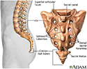
A pelvis x-ray is a picture of the bones around both the hips. The pelvis connects the legs to the body.

How the Test is Performed
The test is done in a radiology department or in the health care provider's office by an x-ray technician.
You will lie down on the table. The pictures are then taken. You may have to move your body to other positions to provide different views.
How to Prepare for the Test
Tell the provider if you are pregnant. Remove all jewelry, especially around your belly and legs. You will wear a hospital gown.
How the Test will Feel
The x-rays are painless. Changing position may cause discomfort.
Why the Test is Performed
The x-ray is used to look for:
- Fractures
- Tumors
- Degenerative conditions of bones in the hips, pelvis, and upper legs
- Abnormal shape of your bones or joint
What Abnormal Results Mean
Abnormal results may suggest:
- Pelvic fractures
- Arthritis of the hip joint
- Tumors of the bones of the pelvis
- Sacroiliitis (inflammation of the area where the sacrum joins the ilium bone)
- Ankylosing spondylitis (abnormal stiffness of the spine and joint)
- Arthritis of the lower spine
- Abnormality of the shape of your pelvis or hip joint

Risks
Children and the fetuses of pregnant women are more sensitive to the risks of the x-ray. A protective shield may be worn over areas not being scanned.
References
Stoneback JW, Gorman MA. Pelvic fractures. In: McIntyre RC, Schulick RD, eds. Surgical Decision Making. 6th ed. Philadelphia, PA: Elsevier; 2020:chap 147.
Williams KD. Spondylolisthesis. In: Azar FM, Beaty JH, eds. Campbell's Operative Orthopaedics. 14th ed. Philadelphia, PA: Elsevier; 2021:chap 40.
Review Date: 6/13/2021
Reviewed By: C. Benjamin Ma, MD, Professor, Chief, Sports Medicine and Shoulder Service, UCSF Department of Orthopaedic Surgery, San Francisco, CA. Also reviewed by David Zieve, MD, MHA, Medical Director, Brenda Conaway, Editorial Director, and the A.D.A.M. Editorial team.
 Todos los derechos son reservados