
Ultrasound pregnancy
Pregnancy sonogram; Obstetric ultrasonography; Obstetric sonogram; Ultrasound - pregnancy; IUGR - ultrasound; Intrauterine growth - ultrasound; Polyhydramnios - ultrasound; Oligohydramnios - ultrasound; Placenta previa - ultrasound; Multiple pregnancy - ultrasound; Vaginal bleeding during pregnancy - ultrasound; Fetal monitoring - ultrasound
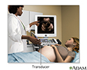
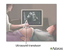
A pregnancy ultrasound is an imaging test that uses sound waves to create a picture of how a baby is developing in the womb. It is also used to check the female pelvic organs during pregnancy.
How the Test is Performed
To have the procedure:
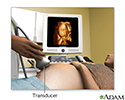
- You will lie on your back on an exam table.
- The person performing the test will spread a clear, water-based gel on your belly and pelvis area. A handheld probe will then be moved over the area. The gel helps the probe transmit sound waves.
- These waves bounce off the body structures, including the developing baby, to create a picture on the ultrasound machine.
In some cases, a pregnancy ultrasound may be done by placing the probe into the vagina. This is more likely in early pregnancy, Many women will have the length of their cervix measured by vaginal ultrasonography around 20 to 24 weeks of pregnancy.

How to Prepare for the Test
You will need to have a full bladder to get the best ultrasound image. You may be asked to drink 2 to 3 glasses of liquid an hour before the test. DO NOT urinate before the procedure.
How the Test will Feel
There may be some discomfort from pressure on the full bladder. The conducting gel may feel slightly cold and wet. You will not feel the ultrasound waves.
Why the Test is Performed
An ultrasound may be done to determine if there is a problem with the pregnancy, how far along the pregnancy is, or to take measurements and screen for potential problems.
Talk to your health care provider to determine the most appropriate scanning schedule for you.
A pregnancy ultrasound may be done during the first 12 weeks of pregnancy to:
- Confirm a normal pregnancy
- Determine the baby's age
- Look for problems, such as ectopic pregnancies or the chances for a miscarriage
- Determine the baby's heart rate
- Look for multiple pregnancies (such as twins and triplets)
- Identify problems of the placenta, uterus, cervix, and ovaries
- Look for findings that might indicate an increased risk for Down syndrome
A pregnancy ultrasound may also be done in the second and third trimesters to:
- Determine the baby's age, growth, position, and sometimes sex.
- Identify any problems with how the fetus is developing.
- Look for twins or triplets. Look at the placenta, amniotic fluid, and pelvis.
Some centers are now performing a pregnancy ultrasound called a nuchal translucency screening test around 9 to 13 weeks of pregnancy. This test is done to look for signs of Down syndrome or other problems in the developing baby. This test is often combined with blood tests to improve the accuracy of results.
How many ultrasounds you will need depends on whether a previous scan or blood test has detected problems that require follow-up testing.
Normal Results
The developing baby, placenta, amniotic fluid, and surrounding structures appear normal for the gestational age.
Note: Normal results may vary slightly. Talk to your doctor about the meaning of your specific test results.
What Abnormal Results Mean
Abnormal ultrasound results may be due to some of the following conditions:
- Birth defects
- Ectopic pregnancy
- Poor growth of a baby while in the mother's womb
- Multiple pregnancies
- Miscarriage
- Problems with the baby's position in the womb
- Problems with the placenta, including placenta previa and placental abruption
- Too little amniotic fluid
- Too much amniotic fluid (polyhydramnios)
- Tumors of pregnancy, including gestational trophoblastic disease
- Other problems with the ovaries, uterus, and remaining pelvic structures


Risks
Current ultrasound techniques appear to be safe. Ultrasound does not involve radiation.
References
Richards DS. Obstetric ultrasound: imaging, dating, growth, and anomaly. In: Landon MB, Galan HL, Jauniaux ERM, et al, eds. Gabbe's Obstetrics: Normal and Problem Pregnancies. 8th ed. Philadelphia, PA: Elsevier; 2021:chap 9.
Wapner RJ, Dugoff L. Prenatal diagnosis of congenital disorders. In: Resnik R, Lockwood CJ, Moore TR, Greene MF, Copel JA, Silver RM, eds. Creasy and Resnik's Maternal-Fetal Medicine: Principles and Practice. 8th ed. Philadelphia, PA: Elsevier; 2019:chap 32.
Wolf RB. Abdominal imaging. In: Resnik R, Lockwood CJ, Moore TR, Greene MF, Copel JA, Silver RM, eds. Creasy and Resnik's Maternal-Fetal Medicine: Principles and Practice. 8th ed. Philadelphia, PA: Elsevier; 2019:chap 26.
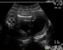
Ultrasound in pregnancy - illustration
Ultrasound in pregnancy
illustration
Ultrasound, normal fetus - abdomen measurements - illustration
Ultrasound, normal fetus - abdomen measurements
illustration
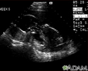
Ultrasound, normal fetus - arm and legs - illustration
Ultrasound, normal fetus - arm and legs
illustration
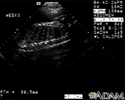
Ultrasound, normal placenta - Braxton Hicks - illustration
Ultrasound, normal placenta - Braxton Hicks
illustration
Ultrasound, normal fetus - face - illustration
Ultrasound, normal fetus - face
illustration
Ultrasound, normal fetus - femur measurement - illustration
Ultrasound, normal fetus - femur measurement
illustration
Ultrasound, normal fetus - foot - illustration
Ultrasound, normal fetus - foot
illustration
Ultrasound, normal fetus - head measurements - illustration
Ultrasound, normal fetus - head measurements
illustration
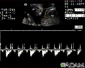
Ultrasound, normal fetus - heartbeat - illustration
Ultrasound, normal fetus - heartbeat
illustration
Ultrasound, ventricular septal defect - heartbeat - illustration
Ultrasound, ventricular septal defect - heartbeat
illustration
Ultrasound, normal fetus - arms and legs - illustration
Ultrasound, normal fetus - arms and legs
illustration
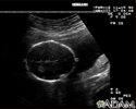
Ultrasound, normal relaxed placenta - illustration
Ultrasound, normal relaxed placenta
illustration
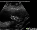
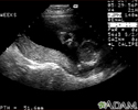
Ultrasound, normal fetus - profile view - illustration
Ultrasound, normal fetus - profile view
illustration
Ultrasound, normal fetus - spine and ribs - illustration
Ultrasound, normal fetus - spine and ribs
illustration
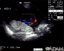
Ultrasound, color - normal umbilical cord - illustration
Ultrasound, color - normal umbilical cord
illustration
Ultrasound, normal fetus - ventricles of brain - illustration
Ultrasound, normal fetus - ventricles of brain
illustration
Prenatal ultrasound - series - Procedure, part 1
Presentation
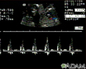
3D ultrasound - illustration
3D ultrasound
illustration


Ultrasound in pregnancy - illustration
Ultrasound in pregnancy
illustration
Ultrasound, normal fetus - abdomen measurements - illustration
Ultrasound, normal fetus - abdomen measurements
illustration
Ultrasound, normal fetus - arm and legs - illustration
Ultrasound, normal fetus - arm and legs
illustration
Ultrasound, normal placenta - Braxton Hicks - illustration
Ultrasound, normal placenta - Braxton Hicks
illustration
Ultrasound, normal fetus - face - illustration
Ultrasound, normal fetus - face
illustration
Ultrasound, normal fetus - femur measurement - illustration
Ultrasound, normal fetus - femur measurement
illustration
Ultrasound, normal fetus - foot - illustration
Ultrasound, normal fetus - foot
illustration
Ultrasound, normal fetus - head measurements - illustration
Ultrasound, normal fetus - head measurements
illustration
Ultrasound, normal fetus - heartbeat - illustration
Ultrasound, normal fetus - heartbeat
illustration
Ultrasound, ventricular septal defect - heartbeat - illustration
Ultrasound, ventricular septal defect - heartbeat
illustration
Ultrasound, normal fetus - arms and legs - illustration
Ultrasound, normal fetus - arms and legs
illustration
Ultrasound, normal relaxed placenta - illustration
Ultrasound, normal relaxed placenta
illustration
Ultrasound, normal fetus - profile view - illustration
Ultrasound, normal fetus - profile view
illustration
Ultrasound, normal fetus - spine and ribs - illustration
Ultrasound, normal fetus - spine and ribs
illustration
Ultrasound, color - normal umbilical cord - illustration
Ultrasound, color - normal umbilical cord
illustration
Ultrasound, normal fetus - ventricles of brain - illustration
Ultrasound, normal fetus - ventricles of brain
illustration
Prenatal ultrasound - series - Procedure, part 1
Presentation
3D ultrasound - illustration
3D ultrasound
illustration
Review Date: 1/10/2022
Reviewed By: John D. Jacobson, MD, Department of Obstetrics and Gynecology, Loma Linda University School of Medicine, Loma Linda, CA. Also reviewed by David Zieve, MD, MHA, Medical Director, Brenda Conaway, Editorial Director, and the A.D.A.M. Editorial team.
 All rights reserved.