
Extremity x-ray
An extremity x-ray is an image of the hands, wrist, feet, ankle, leg, thigh, forearm humerus or upper arm, hip, shoulder or all of these areas. The term "extremity" often refers to a human limb.

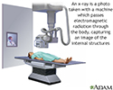
X-rays are a form of radiation that passes through the body to form an image on film. Structures that are dense (such as bone) will appear white. Air will be black, and other structures will be shades of gray.
The test is done in a hospital radiology department or in the health care provider's office. The x-ray is done by an x-ray technologist.
You will need to hold still as the x-ray is taken. You may be asked to change position, so more x-rays can be taken.
How to Prepare for the Test
Tell your provider if you are pregnant. Remove all jewelry from the area being imaged.
In general, there is no discomfort. You may be slightly uncomfortable while the leg or arm is put in place for the x-ray.
Why the Test is Performed
Your provider may order this test if you have signs of:
- A fracture
- Tumor
- Arthritis (inflammation of the joints)
- A foreign body (such as a piece of metal)
- An infection of the bone (osteomyelitis)
- Delayed growth in a child

Normal Results
The x-ray shows normal structures for the age of the person.
What Abnormal Results Mean
Abnormal results may be due to:
- Bone conditions that get worse over time (degenerative)
- Bone tumor
- Broken bone (fracture)
- Dislocated bone
- Osteomyelitis (infection)
- Arthritis
Other conditions for which the test may be performed:
- Clubfoot
- To detect foreign objects in the body

Risks
There is low-level radiation exposure. X-rays are monitored and regulated to provide the smallest amount of radiation exposure needed to make the image. Most experts feel that the risk is low compared with the benefits.
Pregnant women and children are more sensitive to the risks of an x-ray.
References
Kelly DM. Congenital anomalies of the lower extremity. In: Azar FM, Beaty JH, eds. Campbell's Operative Orthopaedics. 14th ed. Philadelphia, PA: Elsevier; 2021:chap 29.
Kim W. Imaging of extremity trauma. In: Torigian DA, Ramchandani P, eds. Radiology Secrets Plus. 4th ed. Philadelphia, PA: Elsevier; 2017:chap 45.
Laoteppitaks C. Compartment syndrome evaluation. In: Roberts JR, Custalow CB, Thomsen TW, eds. Roberts and Hedges' Clinical Procedures in Emergency Medicine and Acute Care. 7th ed. Philadelphia, PA: Elsevier; 2019:chap 54.
Review Date: 7/5/2022
Reviewed By: Jason Levy, MD, FSIR, Northside Radiology Associates, Atlanta, GA. Also reviewed by David C. Dugdale, MD, Medical Director, Brenda Conaway, Editorial Director, and the A.D.A.M. Editorial team.
 All rights reserved.